|
:::
 |
 Now:Home page > Spine Disease > Spinal Fracture Now:Home page > Spine Disease > Spinal Fracture |
|
|
|
 |

|
Example |
 |
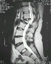
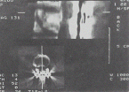
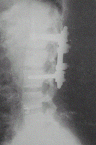
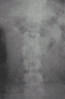
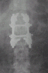
Gender: Male
Age: 22
Symptom Description: A L1 burst fracture was caused by a falling accident, CT scan shows the bone fragments compress the spinal cord and the nerve roots, the patient had neurological deficit and suffered severe pain.
Treatment: Open reduction was performed to restore vertebra to the correct position and instrumentation was used to secure the fracture area. |
|
 |
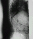
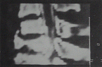
| Before CT |
|
|
 |
| After CT |
|
|
 |
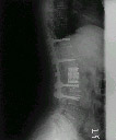
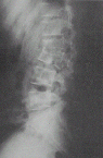
| Before |
|
|
 |
| After |
|
|
 |
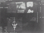
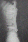
| Before |
|
|
 |
| After |
|
 |
Gender: Male
Age: 24
Symptom Description: The fracture occurs at L2 and L3, that has caused severe pain due to the instability and compression of the nerve root by bone fragments.
Treatment: Open reduction was performed to reduce the fracture and instrumentation was used to secure the correction. |
|
 |
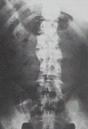
| Before |
|
|
 |
| After |
|
|
 |
| Before |
|
|
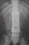 |
| After |
|
 |
Gender: Male
Age: 46
Symptom Description: The injury was caused by the auto accident where the fracture occurs at C7, the bone fragments compressed the spinal cord and cause incomplete paraplegia
Treatment: Corpectomy of C7 was performed to remove the fragments and strut fusion to restore the construct. The neurological function recovered completely. |
|
 |
| Before CT |
|
|
 |
| After CT |
|
 |
Gender: Female
Age: 58
Symptom Description: Old traumatic angular kyphosis at T12. Spinal cord was compressed by retropulsed bony fragment. Pain and neurological deficit were the thief complaint.
Treatment: Combined anterior and posterior procedure were used to decompress the spinal cord, correct the kyphosis and reconstruction the spine. |
|
|
|
|