1spine 1spine 1spine 1spine 1spine 1spine 1spine 1spine 1spine 1spine 1spine 1spine 1spine 1spine 1spine 1spine 1spine 1spine 1spine 1spine 1spine 1spine 1spine 1spine 1spine 1spine 1spine 1spine 1spine 1spine 1spine 1spine 1spine 1spine 1spine 1spine 1spine 1spine 1spine 1spine 1spine 1spine 1spine 1spine 1spine 1spine 1spine 1spine 1spine 1spine 1spine 1spine 1spine 1spine 1spine 1spine 1spine 1spine 1spine 1spine 1spine 1spine 1spine 1spine 1spine 1spine 1spine 1spine 1spine 1spine 1spine 1spine 1spine 1spine 1spine 1spine 1spine 1spine 1spine 1spine 1spine 1spine 1spine 1spine 1spine 1spine 1spine 1scoliosis 1scoliosis 1scoliosis 1scoliosis 1scoliosis 1scoliosis 1scoliosis 1scoliosis 1scoliosis 1scoliosis 1scoliosis 1scoliosis 1scoliosis 1scoliosis 1scoliosis 1scoliosis 1scoliosis 1scoliosis 1scoliosis 1scoliosis 1scoliosis 1scoliosis 1scoliosis 1scoliosis 1scoliosis 1scoliosis 1scoliosis 1scoliosis 1scoliosis 1scoliosis 1scoliosis 1scoliosis 1scoliosis 1scoliosis 1scoliosis 1scoliosis 1scoliosis 1scoliosis 1scoliosis 1scoliosis 1scoliosis 1scoliosis 1scoliosis 1scoliosis 1scoliosis 1scoliosis 1scoliosis 1scoliosis 1scoliosis 1scoliosis 1scoliosis 1scoliosis 1scoliosis 1scoliosis 1scoliosis 1scoliosis 1scoliosis 1scoliosis 1scoliosis 1scoliosis 1scoliosis 1scoliosis 1scoliosis 1kyphosis 1kyphosis 1kyphosis 1kyphosis 1kyphosis 1kyphosis 1kyphosis 1kyphosis 1kyphosis 1kyphosis 1kyphosis 1kyphosis 1kyphosis 1kyphosis 1kyphosis 1kyphosis 1kyphosis 1kyphosis 1kyphosis 1kyphosis 1kyphosis 1kyphosis 1kyphosis 1kyphosis 1kyphosis 1kyphosis 1kyphosis 1kyphosis 1kyphosis 1kyphosis 1kyphosis 1kyphosis 1kyphosis 1kyphosis 1kyphosis 1kyphosis 1kyphosis 1kyphosis 1kyphosis 1kyphosis 1kyphosis 1kyphosis 1kyphosis 1kyphosis 1kyphosis 1kyphosis 1kyphosis 1kyphosis v 1spine surgery 1spine surgery 1spine surgery 1spine surgery 1spine surgery 1spine surgery 1spine surgery 1spine surgery 1spine surgery 1spine surgery 1spine surgery 1spine surgery 1spine surgery 1spine surgery 1spine surgery 1spine surgery 1spine surgery 1spine surgery 1spine surgery 1spine surgery 1spine surgery 1spine surgery 1spine surgery 1spine surgery 1spine surgery 1spine surgery 1spine surgery 1spine surgery 1spine surgery 1spine surgery 1spine surgery 1spine surgery 1spine surgery 1spine surgery 1spine surgery 1spine surgery 1spine surgery 1spine surgery 1spine surgery 1spine surgery 1spine surgery 1spine surgery 1spine surgery 1spine surgery 1spine surgery 1spine surgery 1spine surgery
blank
blank
TaiwanSpineCenter
Print Media
blank
blank
 
blank
blank
 
Ankylosing spondylitis Kyphosis
line
 
Congenital Kyphosis
line
 
Traumatic Kyphosis
line
 
Scheuermann's Kyphosis
line
 
Osteoporotic Kyphosis
line
 
Degenerative Kyphosis
line
 
Iatrogenic Kyphosis
line
 
Postinfectious Kyphosis


blank
:::
blank
icon Now:Home page > Osteoporotic Kyphosis
icon

Back


*

Osteoporotic Kyphosis

*

Approximately 200,000 to 500,000 vertebral compression fractures occur each year in the United States, costing an estimated $250 million. Compression fractures are most common among the elderly, individuals with osteoporosis, and postmenopausal women, occurring in 20%of people over the age of 70 years and in 16% of postmenopausal women. Eighty-four percent of patients with radiographic evidence of compression fracture report associated back pain.

(see figure 1)

*
*
 

Vertebra becomes shorter and leaning forward

Normal
Wedge Fvacture


*

Clinic Study

*


DeSmet et al. did a prospective study of the distribution of thoracic and lumbar spinal fractures and their relationship to thoracic kyphosis in 87 women with osteoporosis. Anterior wedge fractures were noted more commonly in the thoracic spine and the thoracolumbar junction, whereas central compression fracture were more common in the first and fourth lumbar levels. Solitary wedge did not occur above the seventh thoracic vertebral. They concluded that kyphosis in osteoporotic women is significantly related to compression fracture. But other nonskeletal factors contribute as well.

The fractures themselves may also promote further fractures. Wedge fracture of the vertebral are due to those mechanical factors which pass the center of gravity anterior to the vertebral bodies. Anterior wedge fractures increase dorsal and lumbar kyphosis, further shifting the center of gravity, fracture aggravating the tendency for further wedge fractures. Hunch back will affect the normal function of lung and other internal organs due to the compression, it will also severely affect patients health and life expectancy, (see figure 2)

Figure 2: Example of Osteoporotic Kyphosis caused by Multiple Vertebral Compression Fractures.

50 years old
70 years old
*
*
*


*

Treatment

*


The treatment of these fractures is commonly nonoperative and includes bracing and external orthoses, pain medications, physical therapy, and medical therapies for osteoporosis. Many elderly patients, unfortunately have chronic pain and develop progressive kyphotic deformities. Surgical treatment for vertebral compression fractures is indicated only when significant neurologic dysfunction and progressive deformity exist. Such treatment is fraught with complications that are amplified by osteoporosis, including graft dislodgment and subsidence, loss of implant fixation, and junctional kyohosis. Another effective option for the treatment of vertebral compression fractures is vertebroplasty , the percutaneous transpedicular injection of polymrthacylate (PMMA) cement into the vertebral body.

A combined anterior and posterior procedure is always necessary for treatment of progressive severe kyphosis. And it does pose an inordinate risk to senile patients.


*

Abstract

*


Purpose. Current surgical options for osteoporotic kyphosis involve a risk of complex surgery in elderly patients, graft problems (eg, graft dislodgement, subsidence, pseudoarthrosis), and instrumentation problems (eg, adjacent-segment failure, implant pullout). This study is to assess the effectiveness of apical lordosating osteotomy (ALO) and minimal segment fixation through a posterior approach for correcting thoracic or thoracolumbar osteoporotic kyphosis.

Methods. Thirty-two consecutive patients (average age, 71.1 years; range, 65-81 years) with thoracic or thoracolumbar osteoporotic kyphosis underwent ALO. Mean follow-up was 3.2 years (range, 2.1-6.1 years). Radiographic studies, complications, and patient satisfaction were assessed.

Results. Mean operative time was 135 min, and mean blood loss was 722 mL. In 11 patients with thoracic hyperkyphosis, the mean Cobb angle was corrected from 81.6 ° (range, 73 ° -97 °) to 24.3 ° (range, 18 ° -30 °), indicating normal kyphosis. In 19 patients, thoracolumbar kyphosis of 54.4 ° (range, 45°-71°) was corrected to -2.3 ° (range, -11 ° to 8 °). Sagittal imbalance was 11.5 cm before surgery and 4.8 cm after. Satisfactory correction was achieved in all patients, without anterior release. Local kyphosis was corrected to -9.3 ° from 51.2 °, and mean vertebral kyphosis, to -25.3 ° from 15.6 °. In 17 patients with neurologic deficit, Frankel grades improved postoperatively. No major complication occurred. All patients reported improved pain and self-image and overall satisfaction.

Conclusions. ALO and minimal segments fixation is effective for managing thoracic or thoracolumbar osteoporotic kyphosis. It obviated anterior procedures and had fewer complications and high patient satisfaction.

*

Example
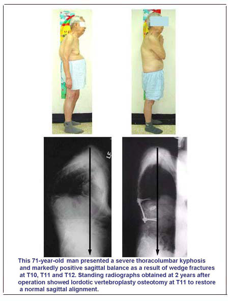
《Press the button to see the surgical example》


blank
  icon

Top

blank
blank