|
:::
 |
|
|
 |

|
Nerve Structure |
| |
 |
| |
Nerves control the body's functions including the vital organs, sensation, and movement. The nervous system receives information and initiates an appropriate response. It is affected by internal and external factors (i.e. stimulus).
Nerves follow tracts and cross over junctions called Synapses. Simplified, it is a complex communicative process between nerves conducted by chemical and/or electrical changes. |
 |
Central Nervous System (CNS) |
| |
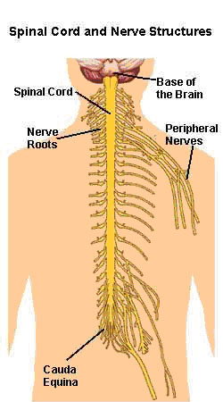
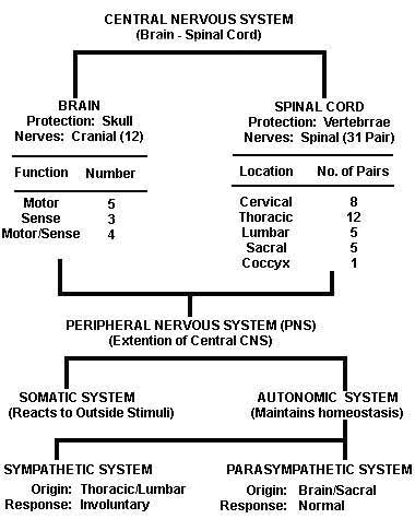
The Central Nervous System is composed of the brain and spinal cord. The brain has 12 Cranial Nerves. The spinal cord, which originates immediately below the brain stem, extends to the first lumbar vertebra (L1). Beyond L1 the spinal cord becomes the Cauda Equina (see below). The spinal cord provides a means of communication between the brain and peripheral nerves.
| BRAIN |
12 Cranial Nerves |
Motor: |
5 nerves |
Sensory: |
3 Nerves |
Motor/Sensory: |
4 nerves |
| SPINAL CORD |
31 Pairs - Spinal Nerves |
Cervical |
8 pair |
Thoracic |
12 pair |
Lumbar |
5 pair |
Sacral |
5 pair |
Coccyx |
1 pair |
|
 |
Peripheral Nervous System (PNS) |
|
| |
 The CNS extends to the Peripheral Nervous System, a system of nerves that branch beyond the spinal cord, brain, and brainstem. The PNS carries information to and from the CNS. The CNS extends to the Peripheral Nervous System, a system of nerves that branch beyond the spinal cord, brain, and brainstem. The PNS carries information to and from the CNS.
The PNS includes the Somatic Nervous System (SNS) and the Autonomic Nervous System (ANS). The somatic nervous system includes the nerves serving the musculoskeletal system and the skin. It is voluntary and reacts to outside stimuli affecting the body. The autonomic nervous system is involuntary automatically seeking to maintain homeostasis or normal function.
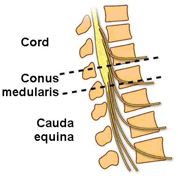
Just below the last Thoracic (T12) and first Lumbar (L1) vertebra the spinal cord ends at the Conus Medullaris. From this point the spinal nerves, resembling a horse's tail become known as the Cauda Equina extending to the coccyx. These nerves are suspended in spinal fluid.
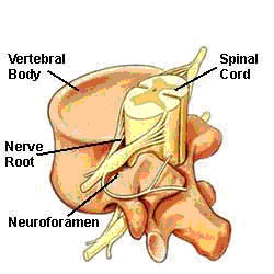
The nerve roots pass out of the spinal canal through the intervertebral foramen, where they feed the body either anteriorly (motor) or posteriorly (sensory). The anterior divisions supply the front of the spine including the limbs. The posterior divisions are distributed to the muscles behind the spine.
|
| |
 |
| |
Spinal Nerves :
(1) Motor:Anterior Roots Ventral Roots
(2) Sensory:Posterior Roots !PDorsal Roots
Other Spinal Cord and Nerve Structures |
 |
Cerebrospinal Fluid (CSF) |
| |
Cerebrospinal fluid is a clear fluid found in the brain chambers (Ventricles), spinal canal, and spinal cord. This fluid is secreted from the Choroids Plexus, a vascular part in the ventricles of the brain. CSF bathes and circulates among these tissues and acts as a shock absorber to protect against injury. The fluid contains different electrolytes, proteins, and glucose. In an average adult the total volume of CSF is about 150 milliliters.
|
 |
Meninges |
| |
Meninges are membranes that cover and protect the brain and spinal cord. There are three primary types: (1) Dura Mater, (2) Arachnoid Mater, and (3) Pia Mater.
- The dura mater, or dura, is the gray outer layer of the spinal cord and nerve roots. It is made of strong connective tissue.
- The arachnoid mater resembles a loosely woven fabric of arteries and veins. This layer is thinner than the dura mater. The Subarachnoid space is filled with cerebrospinal fluid.
- The pia mater is the innermost layer and is a delicate and highly vascular membrane providing blood to the neural structures.
|
|
|
|
|
|